Over the past year, we have established a fantastic relationship with icometrix, a company out of Leuven, Belgium committed to transforming patient care through imaging AI.
The diagnosis of intracranial pathologies with non-contrast computed tomography (NCCT) following TBI is time-sensitive as it involves visual inspection by radiologists and manual quantitative estimation of hematoma size and midline shift. Several limitations of this visual inspection of NCCT include:
- The subjective nature of human radiological interpretation, which is dependent on interpreter experience.
- Well-described considerable inter-center variation and discordance among radiologists when interpreting CT scans for patients with TBI.
- The global shortage of radiologists.
To reduce or eliminate these limitations, icometrix has developed a computer-aided diagnostic system for TBI (icobrain ct), utilizing deep learning (DL) models to increase consistency of CT reads, to reduce diagnostic time lapse, and to provide an objective quantitative evaluation.
icobrain ct is a class II FDA-cleared medical device and is clinically available in the US, Europe, Canada, Australia, Brazil and Japan. icobrain has been validated technically (Jain et al, J Neurotrauma 2019) and through a multi-reader study (Jiang et al. Neuroradiology 2023); however, real-time collection and analysis of evidence for broad clinical adoption/implementation is still to be undertaken.
The icobrain ct output includes:
- Unnormalized measurement of the midline shift, i.e., the shift of the brain past its center line
- Unnormalized volumes of NCCT hyperdensities (epidural, subdural, intraparenchymal and their total); and largest hyperdensity component
- Normalized volumes of the left and right lateral ventricles, and of the fourth ventricle
- Normalized volumes of basal cisterns (suprasellar, quadrigeminal, prepontine & ambient)
- Asymmetry index between the left and right lateral ventricle
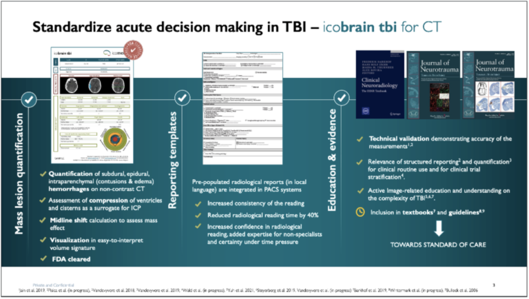
Quantified and automated structured reporting of TBI lesions can provide clinicians with an immediate scan result, inform formal neuroradiological review, and provide objective volumetric information within five minutes. As an example, contusion volume expansion assessed by quantitative image analysis (like icobrain) of head CT scans, might better predict worse functional outcomes.